
Reading a comprehensive hormone panel correctly requires understanding what each marker actually measures, what the optimal range looks like for a performance-oriented man in his prime, how individual markers interact with each other, and critically – what the pattern of results tells you that no single number can. This guide walks through that process systematically.
Step 1: Get the Right Panel Before Interpreting Anything
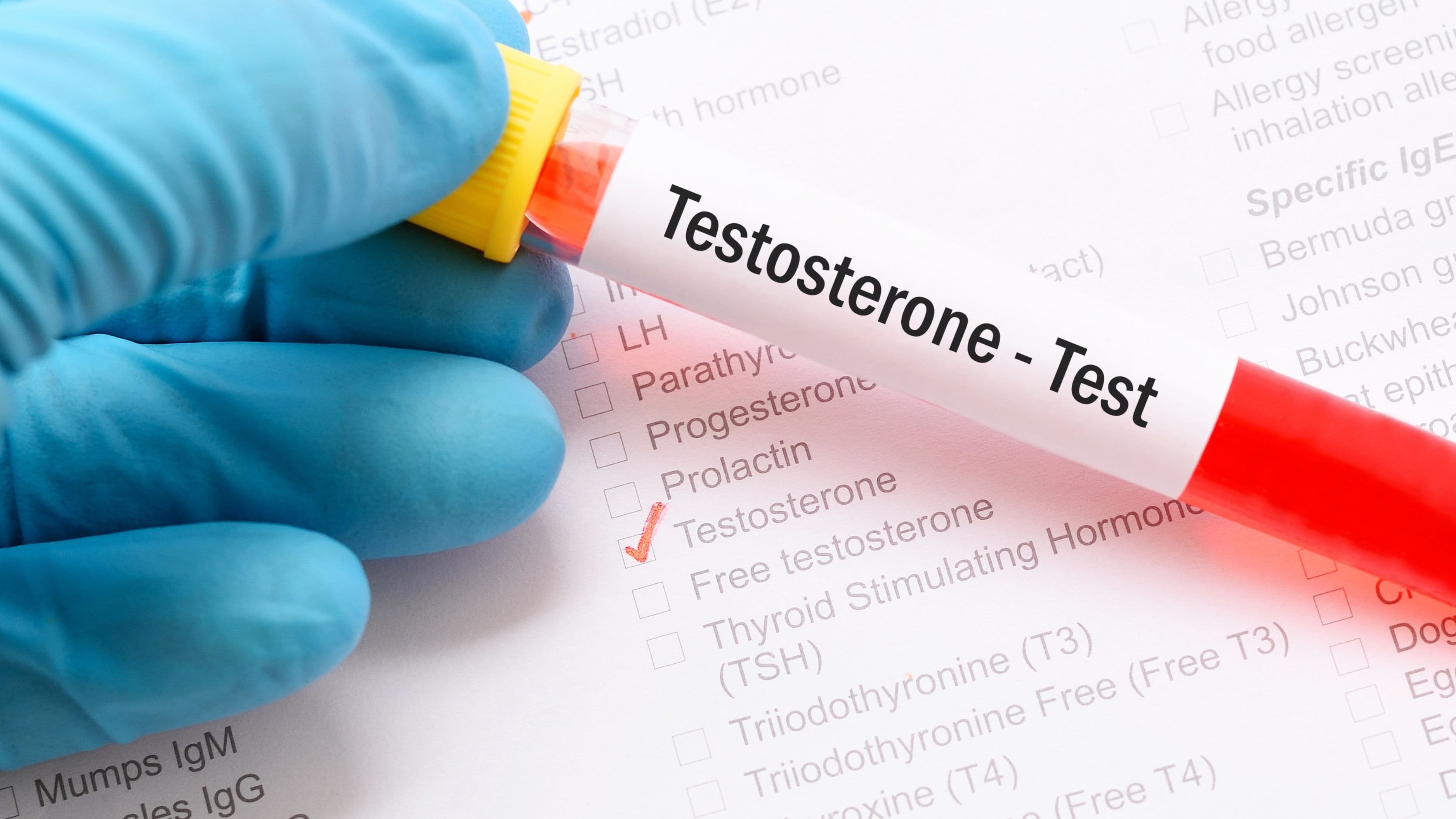
A standard testosterone test ordered by a primary care physician typically measures total testosterone only. That single value is insufficient for any meaningful hormonal assessment. The minimum comprehensive male hormone panel includes the following markers:
Total testosterone, free testosterone, SHBG (Sex Hormone Binding Globulin), LH (luteinizing hormone), FSH (follicle-stimulating hormone), estradiol (E2, sensitive assay), DHEA-S, cortisol (morning draw at minimum, ideally diurnal), prolactin, CBC (complete blood count) for hematocrit and hemoglobin, and a metabolic panel including fasting glucose, insulin, and a lipid panel.
Thyroid function is frequently omitted but directly relevant to hormonal health and should be included: TSH, free T3, free T4, and ideally reverse T3. Men optimizing at the frontier also add IGF-1 (growth hormone proxy), PSA (baseline before age 40, mandatory before initiating TRT), and a diurnal cortisol via DUTCH or four-point salivary testing for a complete adrenal picture.
Timing matters for several markers. Total and free testosterone should be drawn between 7:00–9:00 AM, when levels are at their daily peak. Drawing testosterone at 2:00 PM can show values 20–30% lower than morning levels and produce a false picture of deficiency or, if you're monitoring TRT, an artificially depressed trough reading.
Step 2: Interpret Total Testosterone – But Don't Stop There
Total testosterone is the starting point, not the conclusion. The conventional clinical range runs from approximately 270–1100 ng/dL, a bandwidth wide enough to be nearly meaningless. A 35-year-old man presenting with fatigue, reduced libido, impaired recovery, and declining muscle mass at 310 ng/dL is clinically "normal" by standard criteria. He is not fine.
For a performance-oriented man aged 20–45, an optimal total testosterone range is generally considered to be 600–900 ng/dL. Values below 500 ng/dL warrant further investigation regardless of age. Values above 1000 ng/dL in an untreated man are worth noting but not inherently problematic – some men have constitutively higher production. The key question is whether the level reflects actual bioavailability, which requires the next two markers.
Step 3: Free Testosterone Is the Number That Actually Matters
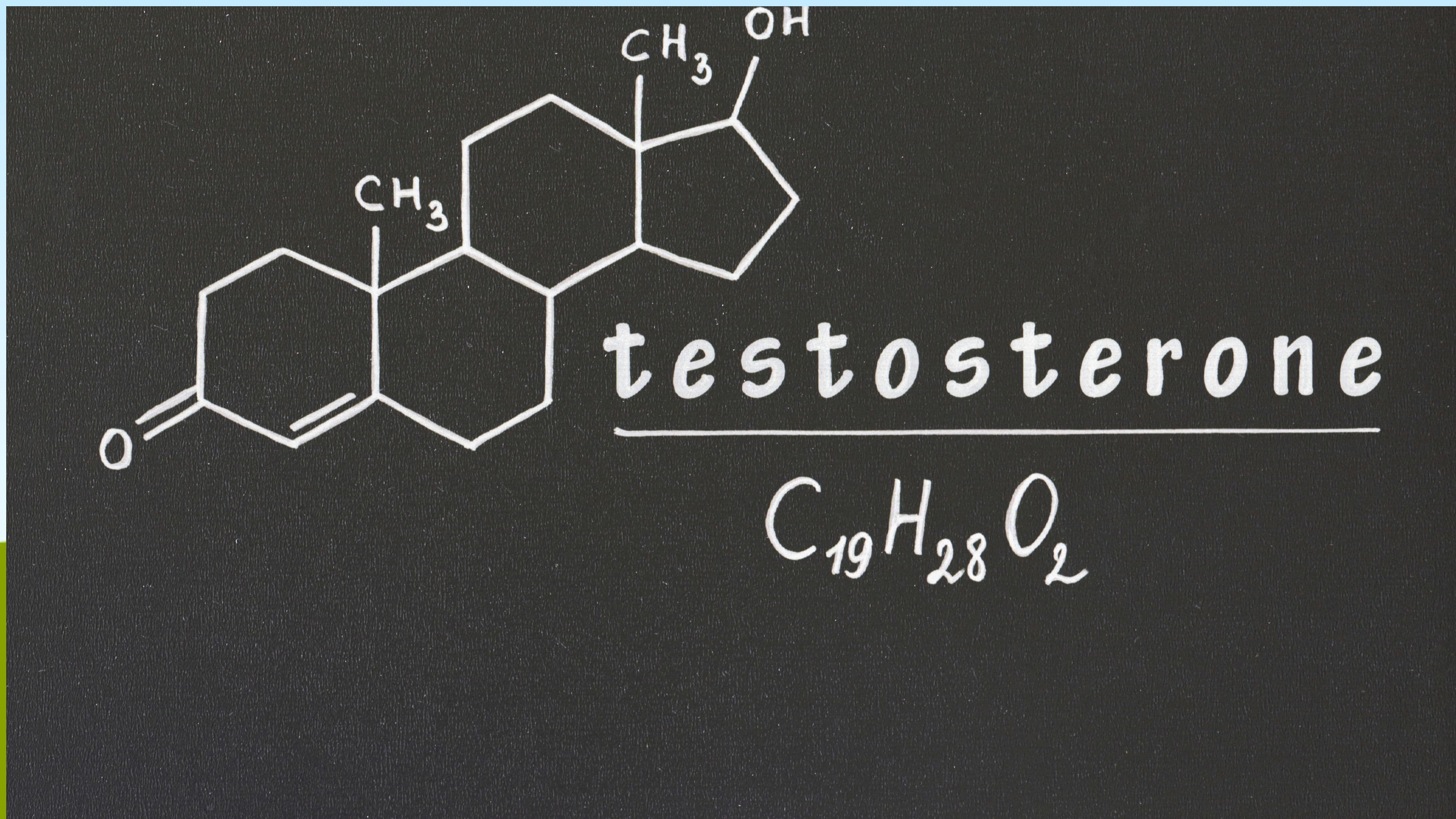
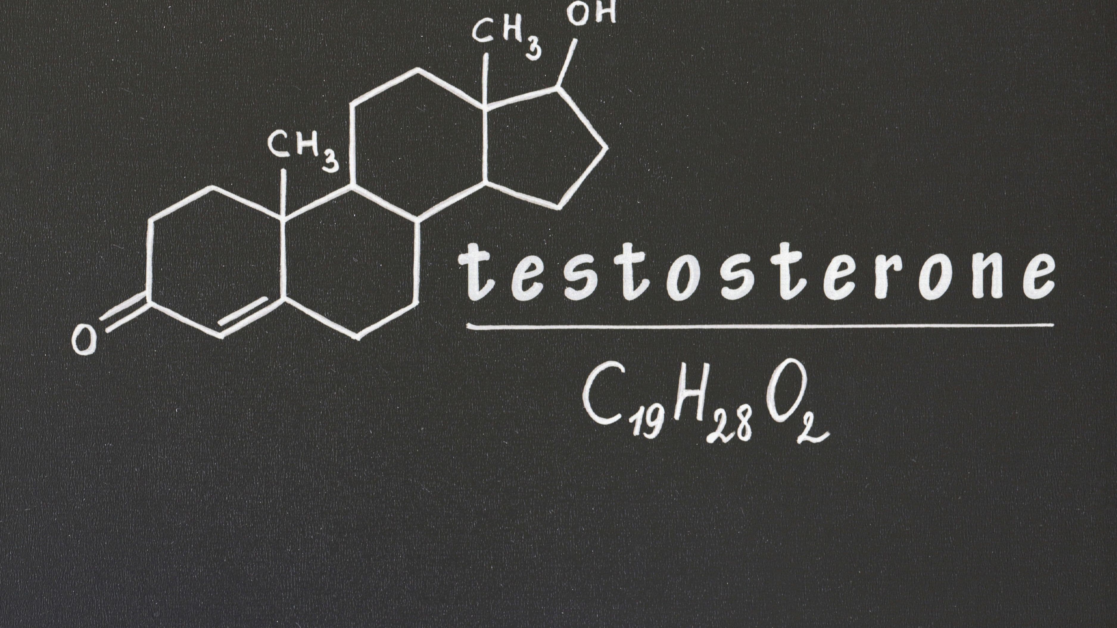
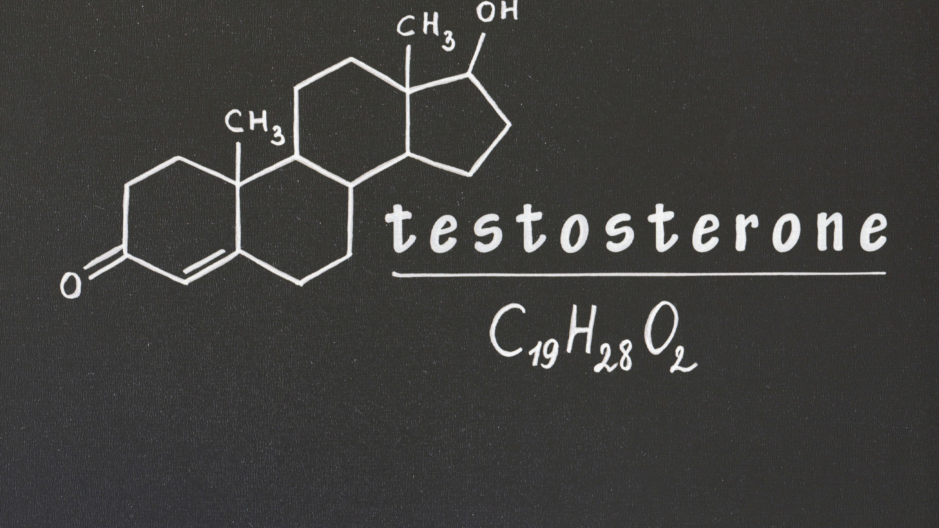
Total testosterone measures all testosterone in circulation – both bound and unbound. The bound fraction, primarily attached to SHBG and albumin, is biologically inactive. Only free testosterone – approximately 1–3% of total – is available to bind androgen receptors and exert physiological effects. Two men with identical total testosterone of 600 ng/dL can have dramatically different androgenic outcomes if their SHBG levels differ substantially.
Free testosterone should be measured directly via equilibrium dialysis, which is the gold standard method. Calculated free testosterone (derived mathematically from total testosterone and SHBG) is an approximation that loses accuracy at the extremes of the SHBG range. Optimal free testosterone is generally considered to be in the range of 15–25 pg/mL (or 150–250 pmol/L depending on assay units), though some men function well at higher or lower values in conjunction with appropriate total testosterone.
If free testosterone is low relative to total testosterone, SHBG elevation is the primary suspect. This shifts the investigation to SHBG and its upstream drivers before any intervention targeting testosterone production itself.
Step 4: Read SHBG as an Independent Hormonal Indicator
SHBG is not merely a carrier protein – it's an independent hormonal actor that responds to multiple metabolic inputs and directly determines testosterone bioavailability. Understanding what drives it up or down is essential for interpreting a panel correctly.
SHBG is elevated by thyroid hormone (both hyperthyroidism and exogenous T3), estrogen, liver pathology (particularly non-alcoholic fatty liver disease in its early stages), low insulin, and aging. It is suppressed by insulin resistance and hyperinsulinemia, anabolic steroids, hypothyroidism, obesity, and chronic low-grade inflammation. The clinical range for SHBG is approximately 10–57 nmol/L. For performance optimization, a target range of 20–35 nmol/L tends to balance adequate free testosterone with the other physiological roles SHBG plays, including its emerging role in cellular signaling independent of testosterone binding.
A man with SHBG at 55 nmol/L and total testosterone at 700 ng/dL may be functionally testosterone-deficient. Before concluding that testosterone production is the problem, his thyroid function, liver enzymes, and insulin sensitivity should all be assessed. Elevated SHBG is often a downstream symptom of a metabolic or thyroid issue, not an isolated finding.
Step 5: LH and FSH Localize the Deficiency
LH and FSH are the pituitary hormones that drive testicular function. They are the diagnostic keys to understanding where in the hormonal cascade a problem originates. Without them, you're looking at outputs without understanding the upstream cause.
LH stimulates Leydig cells to produce testosterone. FSH stimulates Sertoli cells and supports spermatogenesis. Interpreting their levels in conjunction with testosterone reveals the nature of any deficiency.
If total testosterone is low with LH elevated above the reference range (approximately 1.7–8.6 IU/L), this indicates primary hypogonadism – the testes are receiving adequate stimulation but are underperforming. Causes include testicular damage, genetic factors (Klinefelter syndrome), varicocele, or prior steroid-induced Leydig cell desensitization. No lifestyle or supplement protocol corrects primary hypogonadism effectively; TRT is the indicated intervention.
If total testosterone is low with LH low or inappropriately normal – failing to rise in response to low testosterone as it should – this indicates secondary (central) hypogonadism. The problem is upstream: insufficient GnRH signaling from the hypothalamus, insufficient LH output from the pituitary, or suppression from elevated cortisol, hyperprolactinemia, or exogenous androgens. Secondary hypogonadism has more modifiable upstream drivers and is where lifestyle, cortisol management, and in some cases clomiphene or gonadorelin protocols are most relevant.
If LH and FSH are both suppressed with low testosterone, and the man has a history of anabolic steroid use, this is almost certainly HPTA (Hypothalamic-Pituitary-Testicular Axis) suppression from exogenous androgen use. The axis interpretation shifts entirely in this context.
Step 6: Interpret Estradiol Relative to Testosterone, Not in Isolation
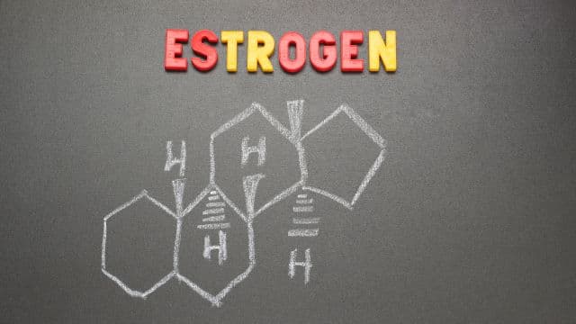
Estradiol in men is produced primarily through peripheral aromatization of testosterone by the enzyme aromatase, with adipose tissue being the dominant conversion site. Some estradiol production also occurs directly in the testes. Estradiol is not the enemy – it plays essential roles in bone density, cardiovascular health, cognitive function, libido, and mood regulation. The problem is excessive estradiol relative to testosterone, not estradiol per se.
Estradiol must be measured using the sensitive assay (sometimes labeled as "estradiol, sensitive" or "LC-MS/MS assay"), not the standard assay calibrated for female hormone ranges. The standard assay is insufficiently sensitive at male estradiol levels and produces unreliable results. Optimal estradiol for men is generally considered to be in the range of 20–30 pg/mL. Values below 15 pg/mL are associated with joint pain, decreased libido, depression, and impaired bone remodeling. Values above 40 pg/mL are associated with water retention, gynecomastia, blunted libido, and mood dysregulation.
The testosterone-to-estradiol ratio is more clinically meaningful than either marker in isolation. A useful target ratio is approximately 20:1 (testosterone in ng/dL divided by estradiol in pg/mL). High aromatase activity – driven primarily by excess adipose tissue, chronic alcohol consumption, insulin resistance, and zinc deficiency – elevates estradiol and suppresses this ratio. Interventions targeting aromatase activity (body composition improvement, zinc repletion, dietary changes) are the first line before pharmaceutical aromatase inhibitors, which carry meaningful risks including joint degradation and bone density loss when overused.
Step 7: Prolactin – Low on the Priority List Until It Isn't
Prolactin is often overlooked in performance-focused panels, but it warrants inclusion because elevated prolactin (hyperprolactinemia) is a direct suppressor of LH pulsatility and testosterone production. The reference range is approximately 2–18 ng/mL for men. Values above 25 ng/mL warrant investigation. Values above 100 ng/mL suggest a pituitary adenoma (prolactinoma) and require imaging.
Modest prolactin elevations (20–40 ng/mL) are common with chronic psychological stress, dopamine-suppressing medications (including some antihistamines and proton pump inhibitors), hypothyroidism, and excessive cannabis use. Dopamine is the primary inhibitory regulator of prolactin secretion; anything that reduces dopaminergic tone will elevate prolactin. If prolactin is mildly elevated and testosterone is suppressed with low-normal LH, this is a meaningful finding that points toward dopaminergic interventions (L-tyrosine, vitamin B6 in P5P form, mucuna pruriens) or removal of the suppressive factor before more aggressive testosterone interventions.
Step 8: Thyroid Markers – The Most Commonly Missed Testosterone Suppressor
TSH alone is insufficient for thyroid assessment. A man can have normal TSH with impaired thyroid hormone conversion, producing a functional hypothyroid state that suppresses testosterone through multiple pathways: SHBG elevation, impaired Leydig cell function, reduced LH pulsatility, and direct metabolic effects on steroidogenesis.
Optimal TSH for performance purposes is 1.0–2.0 mIU/L, tighter than the clinical range of 0.4–4.0 mIU/L. Free T3 is the active thyroid hormone that drives metabolic effects at the cellular level; an optimal range is 3.5–4.0 pg/mL. Free T4 should be in the upper half of the reference range (approximately 1.2–1.8 ng/dL). Elevated reverse T3 – an inactive metabolite that competes with free T3 for receptor binding – indicates stress, caloric restriction, or illness suppressing active thyroid hormone availability and should be assessed if free T3 is low with normal T4.
If TSH is above 2.5 mIU/L or free T3 is below 3.2 pg/mL in a man with low testosterone, resolving the thyroid issue often produces secondary improvement in testosterone and SHBG without any direct testosterone intervention. The sequence matters: address thyroid first, retest testosterone at 8 weeks.
Step 9: Cortisol and DHEA-S – The Adrenal Picture
A morning serum cortisol between 15–25 mcg/dL is the expected range for a man without adrenal dysfunction. Values below 10 mcg/dL suggest adrenal insufficiency or HPA burnout and warrant further evaluation. Values consistently above 25–30 mcg/dL indicate chronic HPA activation and will suppress the HPG axis through the mechanisms detailed in the cortisol-testosterone relationship.
DHEA-S (the sulfated storage form of DHEA, produced by the adrenal glands) is a meaningful longevity and hormonal health marker. Peak DHEA-S in men occurs around age 25–30 and declines approximately 2% per year thereafter. Optimal range for performance is approximately 350–500 mcg/dL. Values below 200 mcg/dL in men under 45 indicate significant adrenal reserve depletion. DHEA serves as a precursor to both testosterone and estrogen and has direct effects on brain function, immune regulation, and cortisol buffering. DHEA supplementation at 25–50 mg/day can be appropriate in confirmed deficiency, but should be monitored for downstream conversion to estrogen, particularly in men with elevated aromatase activity.
Step 10: Build the Pattern, Not the Symptom List
Reading a hormone panel correctly means synthesizing the pattern across markers, not reacting to individual out-of-range values in isolation. A few common patterns and what they indicate:
Low total testosterone + high LH + normal SHBG points to primary testicular insufficiency. The pituitary is doing its job; the testes are not. TRT is likely indicated after ruling out reversible causes.
Low total testosterone + low LH + normal prolactin + elevated cortisol points to HPA-driven central suppression. Address cortisol, sleep, and psychological stress load first. Retest in 8–12 weeks before any androgen intervention.
Normal total testosterone + high SHBG + low free testosterone + elevated TSH points to thyroid-driven SHBG elevation as the primary mechanism. Thyroid optimization is the intervention, not testosterone support.
Normal total testosterone + low SHBG + elevated estradiol + insulin resistance points to metabolic-driven aromatase excess. Body composition improvement and insulin sensitivity are the levers. Aromatase inhibitors without addressing the metabolic root cause will produce incomplete and temporary results.
Low testosterone + low LH + low FSH + history of anabolic steroid use points to HPTA suppression. PCT (post-cycle therapy) protocols or TRT depending on severity and recovery timeline.
Common Mistakes When Acting on Panel Results
Treating a single marker without understanding the pattern is the most common and most costly error. Men who see low testosterone and immediately seek TRT without assessing LH, FSH, thyroid, cortisol, and metabolic markers miss reversible causes and may initiate an exogenous hormone protocol they didn't need.
Relying on standard reference ranges rather than optimal performance ranges produces chronic underdiagnosis. A total testosterone of 350 ng/dL is "normal" by clinical standards. It is not compatible with optimized performance, body composition, or cognitive function in a man under 50.
Testing at the wrong time of day distorts results. Total testosterone drawn in the afternoon, or after a night of poor sleep, significantly underestimates true baseline. Always test fasted in the morning after a normal night of sleep, away from intense exercise in the preceding 24 hours.
Single-point testing without trend data limits interpretation. Hormone levels fluctuate day to day. A comprehensive baseline panel followed by a retest at 8–12 weeks after an intervention provides the trend data needed to evaluate whether anything is actually changing.
FAQ
How often should I run a comprehensive hormone panel? For a baseline assessment with no active intervention: annually is sufficient. If you're managing an active intervention – whether lifestyle changes, supplementation, or TRT – testing at 6–8 weeks after initiating the change, then quarterly once stable, provides the resolution needed to track and adjust.
Is the DUTCH test a replacement for standard bloodwork? No. DUTCH testing provides diurnal cortisol pattern, cortisol and androgen metabolites, and estrogen metabolism pathways – information that serum testing cannot provide. But it doesn't replace serum measurement of total testosterone, free testosterone, LH, FSH, or SHBG, which require blood draws. They are complementary panels.
What's the best free testosterone assay? Equilibrium dialysis is the gold standard. Calculated free testosterone (derived from total testosterone and SHBG using the Vermeulen formula) is a reasonable approximation at mid-range SHBG values but becomes less accurate at extremes. If SHBG is above 50 or below 15 nmol/L, request direct measurement rather than calculated.
Should I test on a training day? No. Intense exercise acutely elevates cortisol and can transiently suppress or elevate testosterone depending on the timing and intensity. Test on a rest day or at least 24 hours after any significant training session for a clean baseline.
Does cholesterol level affect testosterone production? Yes, directly. Cholesterol is the precursor molecule for all steroid hormones, including testosterone. Aggressively low LDL from statin use or very low dietary fat intake can impair steroidogenesis. If total cholesterol is below 130 mg/dL in a man with low testosterone, dietary fat and cholesterol intake and statin dose (if applicable) are worth investigating as contributing factors.
A hormone panel is a diagnostic tool, not a destination. The value it provides is proportional to how intelligently you read the pattern, how correctly you time the test, and how disciplined you are in addressing root causes rather than chasing individual numbers. Get the right panel, draw it at the right time, read it as a system, and build your intervention from the pattern. That sequence produces results that reactive, single-marker thinking never will.
📚 Sources
Bhasin S et al. – Testosterone Therapy in Men with Hypogonadism: An Endocrine Society Clinical Practice Guideline. Journal of Clinical Endocrinology & Metabolism, 2018: https://academic.oup.com/jcem/article/103/5/1715/4939465
Vermeulen A et al. – A critical evaluation of simple methods for the estimation of free testosterone in serum. Journal of Clinical Endocrinology & Metabolism, 1999: https://pubmed.ncbi.nlm.nih.gov/10523012
Travison TG et al. – The relative contributions of aging, health, and lifestyle factors to serum testosterone decline in men. Journal of Clinical Endocrinology & Metabolism, 2007: https://pubmed.ncbi.nlm.nih.gov/17090633
Hampl R, Stárka L – SHBG – more than a sex hormone carrier. Prague Medical Report, 2009: https://pubmed.ncbi.nlm.nih.gov/19685710
Winters SJ, Kelley DE, Goodpaster B – The analog free testosterone assay: are the results in men clinically useful? Clinical Chemistry, 1998: https://pubmed.ncbi.nlm.nih.gov/9484227
Melmed S et al. – Diagnosis and Treatment of Hyperprolactinemia: An Endocrine Society Clinical Practice Guideline. Journal of Clinical Endocrinology & Metabolism, 2011: https://academic.oup.com/jcem/article/96/2/273/2597012
Garber JR et al. – Clinical Practice Guidelines for Hypothyroidism in Adults (ATA/AACE). Endocrine Practice, 2012: https://pubmed.ncbi.nlm.nih.gov/23246686
Ohlsson C et al. – Low serum testosterone is associated with worse metabolic profile and increased mortality risk in older men. Archives of Internal Medicine, 2011: https://pubmed.ncbi.nlm.nih.gov/21987192